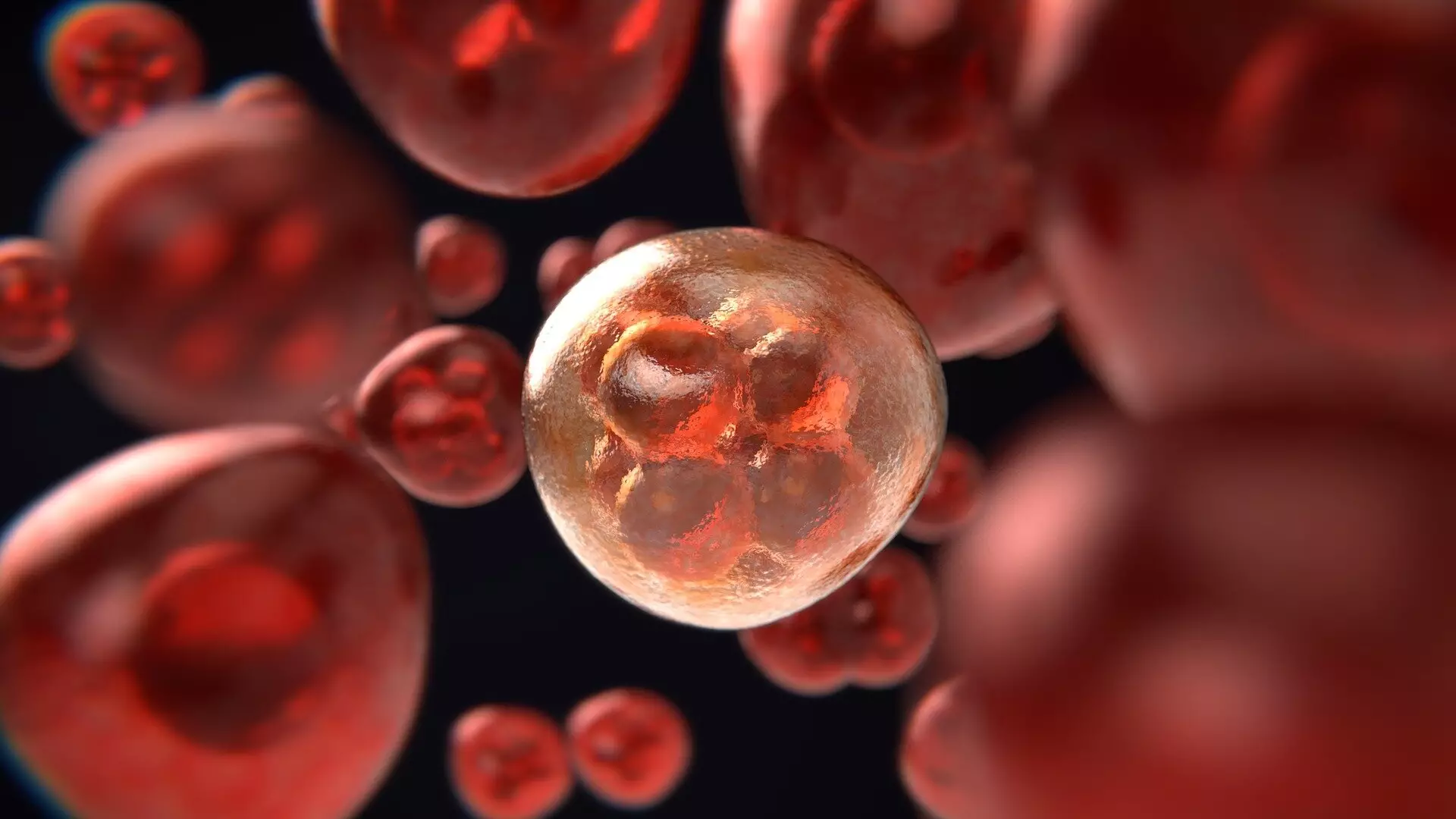
The ability to examine the inner workings of a single cancer cell and how it interacts with surrounding cells is a groundbreaking development in the field of cancer research. Scientists have recently celebrated the advent of a new technique that enables the analysis of the fatty components of cancer cells on an individual basis. Through a collaborative study led by the University of Surrey, researchers have successfully isolated and examined single live cancer cells to determine the lipid compositions within them. This innovative approach, detailed in the journal Analytical Chemistry, offers valuable insights into how cancer cells respond to environmental changes.
Dr. Johanna Von Gerichten, a prominent figure in the study from Surrey’s School of Chemistry and Chemical Engineering, highlighted the significant challenge of cancer cell heterogeneity. Each cancer cell exhibits unique characteristics, making it challenging to devise effective treatment strategies. Traditional methods of studying isolated cells have often been limited in providing detailed analyses of cellular components, hindering a comprehensive understanding of cancer cell biology. The ability to directly sample live cancer cells under a microscope and analyze their lipid contents individually represents a major advancement in cancer research.
Experimental Techniques
The experimental process involved the extraction of individual pancreatic cancer cells from a glass culture dish using Yokogawa’s Single Cellome System SS2000. This state-of-the-art system employs miniature tubes with a diameter of 10 µm, allowing for precise isolation of single live cells. Through the use of fluorescent dye staining, researchers were able to track lipid droplets within the cells throughout the experiment. By collaborating with partners at Sciex, a novel method utilizing a mass spectrometer was developed to fragment the lipids in the cells, providing valuable insights into their composition. The researchers observed distinct lipid profiles among different cells and noted changes in lipid content in response to external stimuli.
Professor Melanie Bailey expressed enthusiasm about the prospects of applying this technique to various cell types beyond cancer cells. The establishment of the national facility for single and sub-cellular “omics,” SEISMIC, will enable further exploration of infection, immunity, and other biological processes. Additionally, participation in the International Atomic Energy Agency program will facilitate the study of the effects of irradiation on cells and the mechanisms underlying resistance to radiation therapy. Dr. Carla Newman, an Associate Director at GSK, emphasized the potential of this innovative method in gaining unprecedented insights into cancer cell communication and paving the way for the development of targeted treatments.
The ability to scrutinize individual cancer cells at a molecular level marks a significant milestone in cancer research. By evaluating the lipid contents of live cells in real-time, scientists have gained valuable knowledge about the dynamic nature of cancer cell metabolism and response to environmental cues. This groundbreaking technique opens up new possibilities for studying various cellular processes and developing customized treatment approaches for cancer and other diseases. The collaborative effort between academic institutions, research organizations, and industry partners underscores the transformative impact of innovative technologies in advancing our understanding of complex biological systems.
Leave a Reply