The field of microscopy has long been a vital tool in the life sciences, allowing biologists to study the intricate details of biological samples at a cellular level. However, one of the challenges faced by microscopists is obtaining clear and sharp images, especially when working with thick biological samples that bend light and create distortions. Traditional adaptive optics techniques have been complex, expensive, and slow, making them out of reach for many research labs.
A team of researchers at HHMI’s Janelia Research Campus has taken a groundbreaking approach by adapting techniques used in astronomy to unblur images of far-away galaxies for use in the life sciences. Astronomers have long employed methods that measure how light is distorted by the atmosphere to correct aberrations and improve the clarity and sharpness of their telescope images.
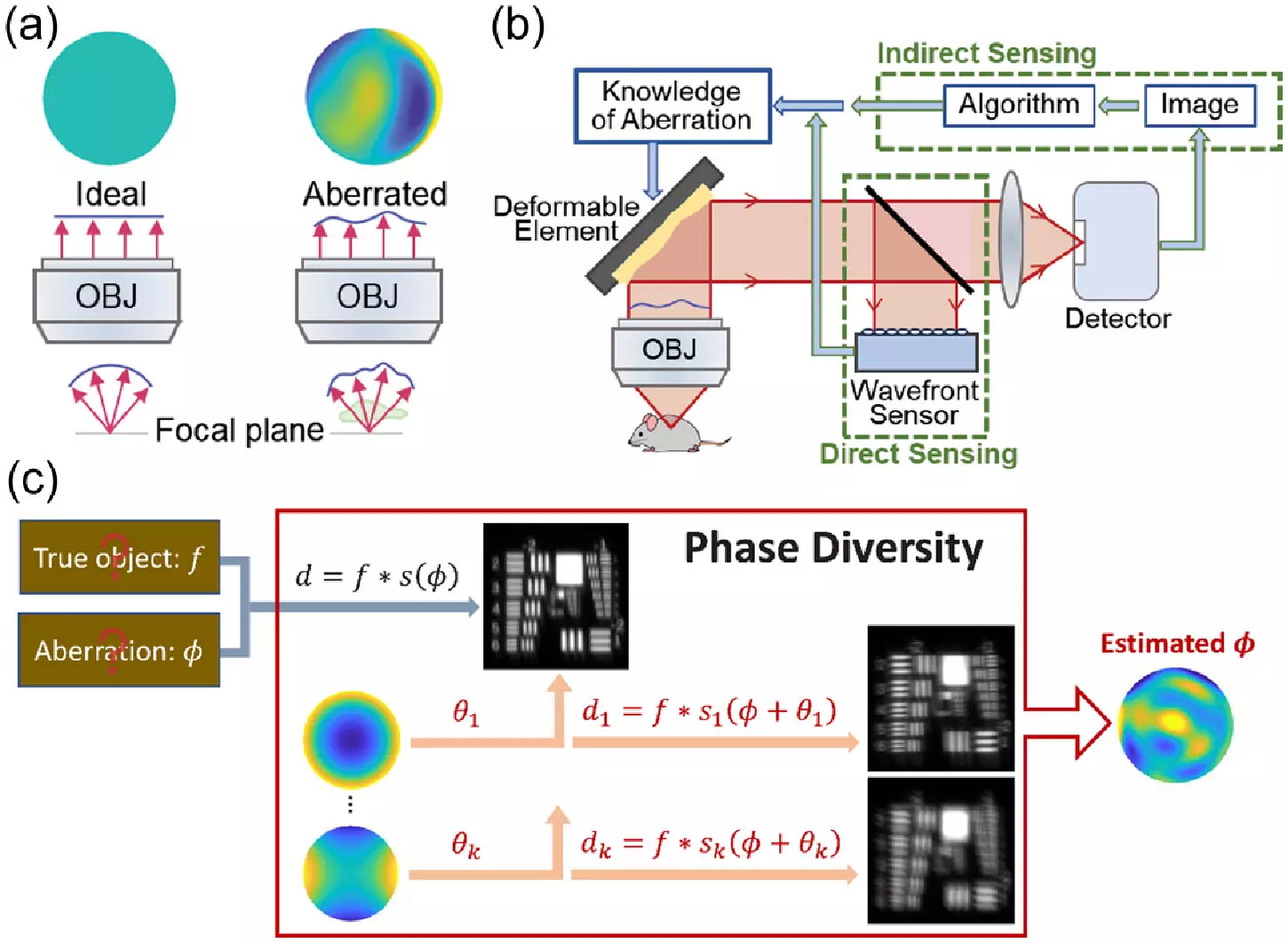
The team at HHMI’s Janelia Research Campus focused on a class of techniques called phase diversity, which has been widely used in astronomy but is relatively new to the life sciences. These methods involve adding additional images with known aberrations to a blurry image with unknown aberrations, providing crucial information to unblur the original image. What sets phase diversity apart is that it does not necessitate major changes to an imaging system, making it an attractive option for microscopy applications.
To implement the new method, the researchers first adapted the astronomy algorithm for microscopy and validated it through simulations. They then constructed a microscope equipped with a deformable mirror, two additional lenses, and improved software to carry out the phase diversity correction. This setup allowed them to calibrate the deformable mirror and correct randomly generated aberrations significantly faster than with existing methods.
The team demonstrated the effectiveness of the new method by obtaining clearer images of fluorescent beads and fixed cells. Their next goal is to test the technique on real-world samples, such as living cells and tissues, and expand its use to more complex microscopes. Additionally, they aim to streamline the process, making it more automated and user-friendly for biologists.
The innovative approach developed by the team at HHMI’s Janelia Research Campus offers a faster and more cost-effective way to implement adaptive optics in microscopy. By making adaptive optics more accessible to a wider range of research labs, biologists will have the opportunity to visualize cellular structures with greater clarity and detail. This advancement has the potential to revolutionize the field of microscopy and pave the way for new discoveries in the life sciences.
Leave a Reply