In recent groundbreaking research, Swansea University scientists have developed a new imaging method for neutral atomic beam microscopes that has the potential to significantly accelerate the process of obtaining microscope images. This innovation could have far-reaching implications for engineers and scientists who rely on microscope imaging for their research and experiments.
Neutral atomic beam microscopes are currently a topic of intense research interest due to their ability to image surfaces that are not easily studied using conventional microscopes. Delicate samples such as bacterial biofilms, ice films, and organic photovoltaic devices present challenges for traditional imaging methods, as they are often damaged or altered by electrons, ions, and photons.
Traditional neutral atomic beam microscopes operate by scanning a microscopic pinhole over the sample, measuring the scattered beam to construct an image pixel by pixel. However, this approach is time-consuming, as each pixel must be measured individually. Attempts to improve resolution by reducing the pinhole size lead to a significant reduction in beam flux, resulting in even longer measurement times.
The Swansea University research team, led by Professor Gil Alexandrowicz from the chemistry department, has introduced a novel and faster method of imaging using neutral atomic beam microscopes. This new technique involves passing a beam of helium-3 atoms through a non-uniform magnetic field, utilizing nuclear spin precession to encode the particles’ positions as they interact with the sample.
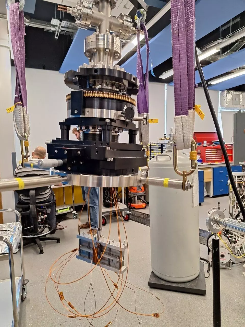
Ph.D. student Morgan Lowe, a member of the Swansea team, constructed the magnetic encoding device and conducted initial experiments to validate the effectiveness of the new method. The beam profile measurements obtained by Mr. Lowe closely matched the results of numerical simulations, demonstrating the accuracy and reliability of the new imaging technique.
Numerical simulations conducted by the research team indicate that the magnetic encoding method has the potential to enhance image resolution without significantly increasing measurement times, in contrast to traditional pinhole scanning methods. Professor Alexandrowicz believes that this new approach opens up exciting possibilities for improving neutral beam microscopy and enabling new contrast mechanisms based on the magnetic properties of samples.
Moving forward, the Swansea University research team plans to further develop the magnetic encoding method to create a fully functional prototype of a magnetic encoding neutral beam microscope. This prototype will allow for comprehensive testing of the technique’s resolution limits, contrast mechanisms, and operational capabilities, paving the way for widespread adoption of this innovative imaging technology.
The research conducted by the Swansea University team represents a significant advancement in the field of neutral atomic beam microscopy. By overcoming the limitations of traditional imaging methods, this new technique has the potential to revolutionize the way scientists and engineers obtain high-resolution microscope images, opening up new possibilities for research and discovery.


Leave a Reply