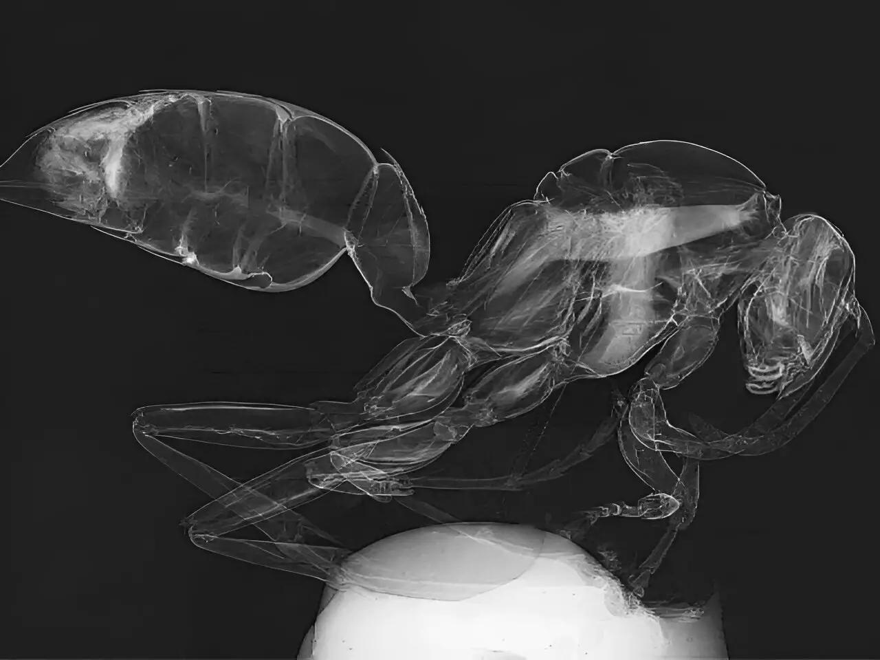
In a recent study conducted at the University of Houston, researchers have introduced a groundbreaking advancement in X-ray imaging technology that has the potential to revolutionize various industries. This innovative approach to X-ray imaging offers significant improvements in medical diagnostics, materials, industrial imaging, transportation security, and other applications. The research, featured on the cover of Optica, highlights the breakthrough achieved by Mini Das, a Moores professor at UH’s College of Natural Sciences and Mathematics, and Jingcheng Yuan, a physics graduate student at UH.
X-ray phase contrast imaging (PCI) has gained considerable attention in recent years due to its ability to provide enhanced contrast for soft tissues by utilizing relative phase changes as X-rays pass through an object. Traditional X-ray imaging methods rely on X-ray absorption to produce an image, which often leads to low contrast and difficulty in distinguishing between materials of similar density. This limitation has been a significant challenge across various fields, including medical imaging and explosive detection.
Das and Yuan introduce a novel light transport model for a single-mask phase imaging system that enhances non-destructive deep imaging for improved visibility of light-element materials, such as soft tissues like cancers, and background tissues like plastics and explosives. The design uses an X-ray mask with periodic slits, creating a compact setup that enhances edge contrast and captures differential phase information to show variations between materials more clearly. One of the main advantages of this system is its simplicity and efficiency, allowing for single-shot, low-dose imaging without the need for high-resolution detectors or complex, multi-shot processes.
The new light transport model developed by Das and Yuan enables a better understanding of contrast formation and how multiple contrast features interact in acquired data. This advancement allows for the retrieval of images with two distinct types of contrast mechanisms from a single exposure, a significant improvement over traditional methods. The simplicity and effectiveness of the single-mask phase imaging system make it a versatile solution for a wide range of imaging challenges in medical diagnostics, materials science, security screening, and other applications.
Das and her team have already tested the model through rigorous simulations and on their in-house developed laboratory benchtop X-ray imaging system. The next step is to integrate this technology into portable systems and retrofit existing imaging setups to test it in real-world environments, such as hospitals, industrial X-ray imaging, and airports. This research paves the way for new possibilities in X-ray imaging by providing a simple, effective, and low-cost method for enhancing image contrast, addressing a critical need for non-destructive deep imaging and improving diagnostics and security screening.
The advancement in X-ray imaging technology by the researchers at the University of Houston represents a significant step forward in the field of medical diagnostics, materials science, industrial imaging, and transportation security. This innovative approach to X-ray imaging offers a simple, effective, and low-cost solution for enhancing image contrast and has the potential to transform the way we approach imaging challenges in various industries. The future looks bright for X-ray imaging with this groundbreaking advancement.
Leave a Reply