The world of microscopy has always been a fascinating realm where scientists delve deep into the unseen structures of living organisms. However, one of the major limitations faced by scientists has been the low resolution of mid-infrared microscopy in comparison to other advanced techniques. While super-resolution fluorescent microscopes and electron microscopes have been able to provide detailed images, they come with their own set of limitations such as sample labeling requirements and the need for samples to be placed in a vacuum, respectively.
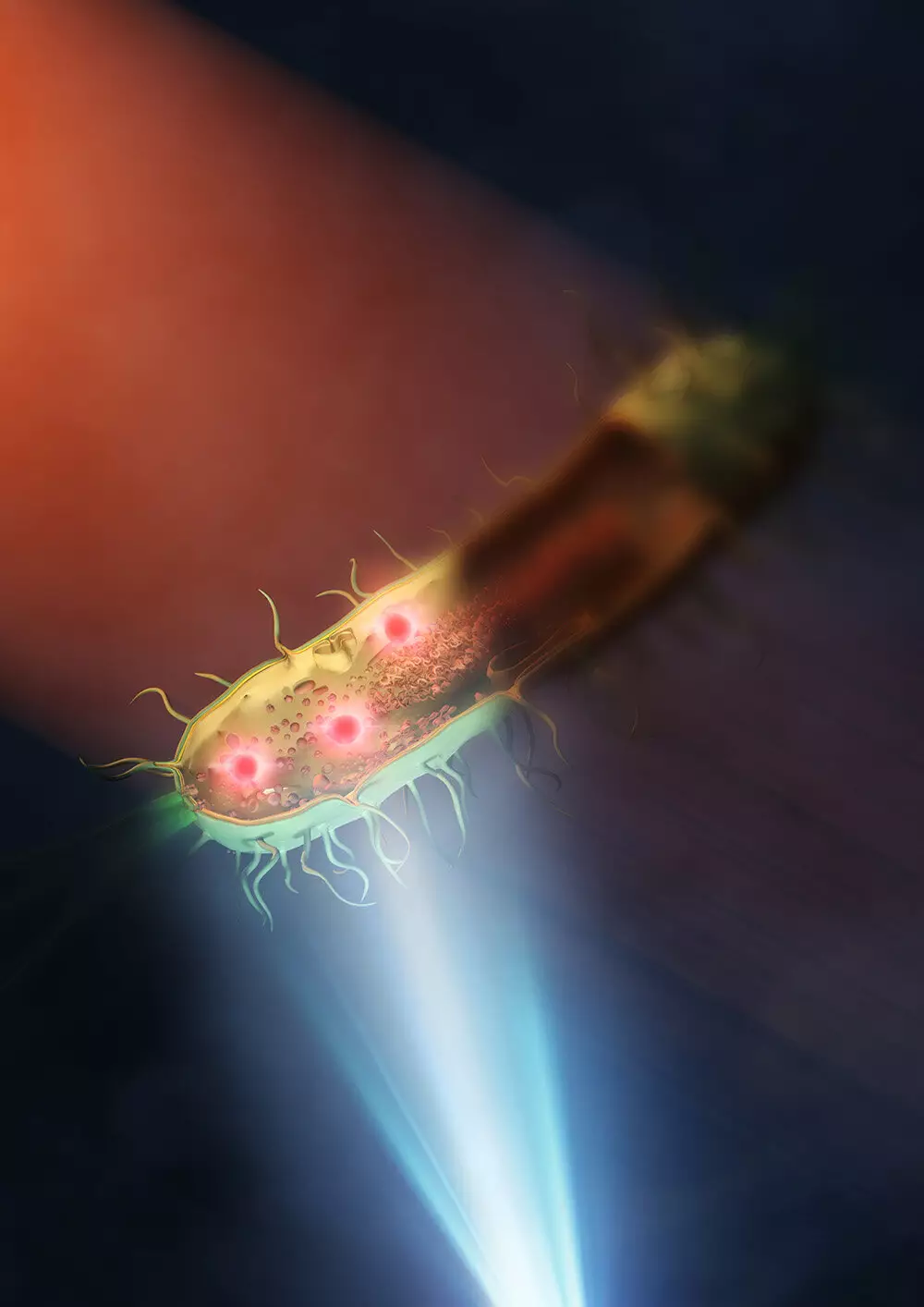
Recently, a team at the University of Tokyo has made a significant advancement in the field of mid-infrared microscopy. They have successfully created an improved mid-infrared microscope that allows them to see the intricate structures inside living bacteria at a nanometer scale. This breakthrough has been published in Nature Photonics and marks a 30-fold improvement in the resolution capabilities of typical mid-infrared microscopes.
The key to achieving this remarkable feat lies in the use of a “synthetic aperture” technique that combines multiple images taken from different illuminated angles to create a clearer overall picture. By placing the sample, which in this case was bacteria (E. coli and Rhodococcus jostii RHA1), on a silicon plate that reflected visible light and transmitted infrared light, the researchers were able to overcome the limitations posed by traditional lenses. This innovative approach allowed for better illumination of the sample with mid-infrared light, resulting in highly detailed images with a spatial resolution of 120 nanometers.
The ability to view live cells at such a small scale opens up a myriad of possibilities for various fields of research, including infectious diseases and antimicrobial resistance. The newfound capability to provide both chemical and structural information about cells, without the need for labeling or damaging samples, is a significant advantage in biological research.
Professor Takuro Ideguchi from the Institute for Photon Science and Technology at the University of Tokyo, who led the research team, expressed optimism about the future potential of this technique. He mentioned that by utilizing better lenses and shorter wavelengths of visible light, the spatial resolution could potentially be further improved to below 100 nanometers. This holds promise for the development of even more accurate mid-infrared-based imaging in the future.
The breakthrough achieved by the University of Tokyo research team in enhancing the resolution of mid-infrared microscopy opens up new possibilities for studying the intricate structures of living organisms at a nanometer scale. This advancement could pave the way for groundbreaking discoveries in various fields of research and propel the field of microscopy to new heights.
Leave a Reply