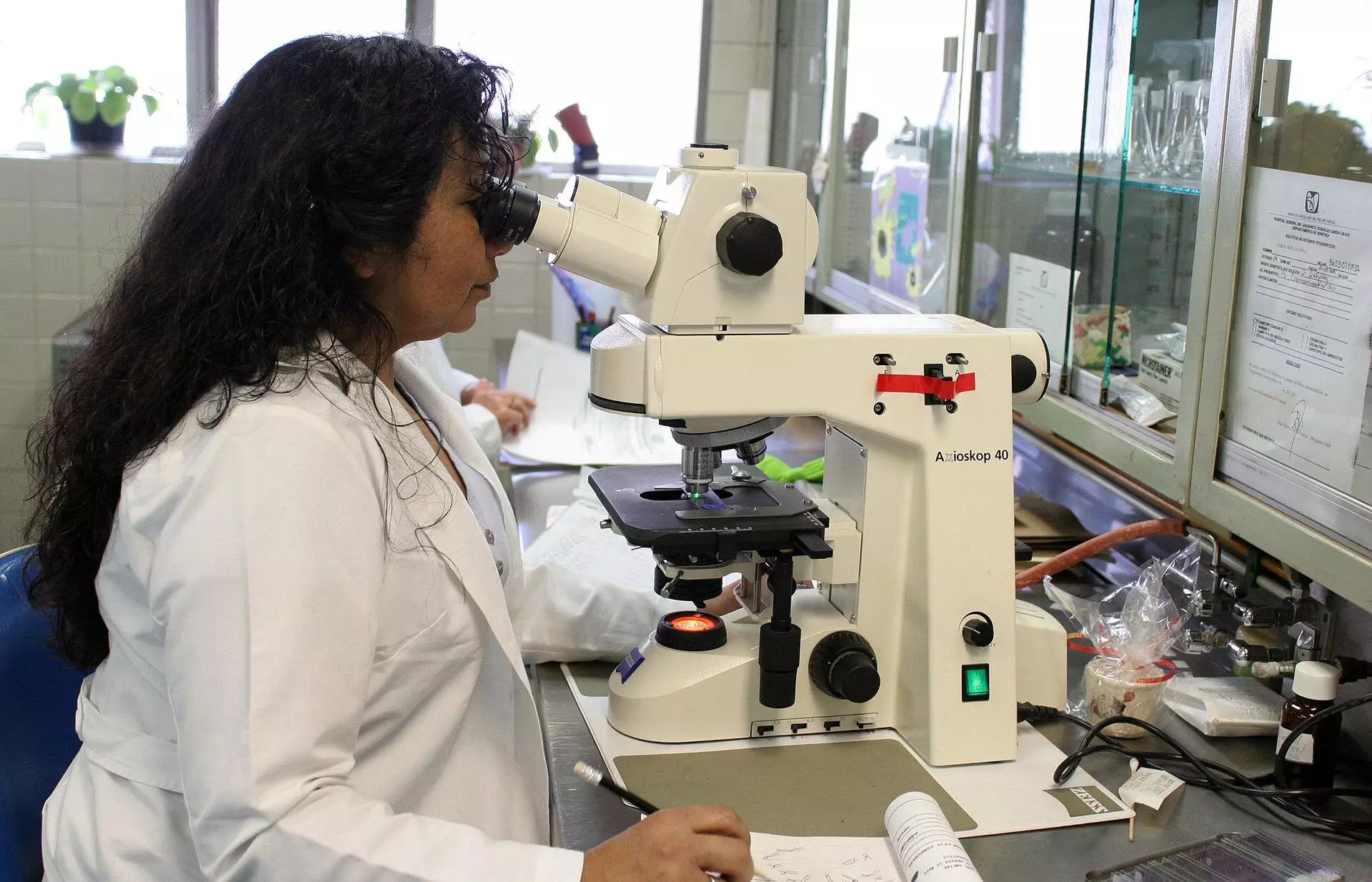
When using a microscope to view biological samples, it is crucial to consider the potential for distortion caused by the difference in refractive indices between the lens medium and the sample medium. This phenomenon results in the sample appearing flattened due to the bending of light rays. In the past, it was assumed that the corrective factor for this distortion was constant regardless of the sample depth.
Recent research conducted by Sergey Loginov, Daan Boltje, and Ernest van der Wee has shed light on the depth-dependent nature of this flattening effect. Through calculations and a mathematical model, it was demonstrated that the sample appears more strongly flattened closer to the lens compared to farther away. This discovery challenges the previously held belief of a constant corrective factor and emphasizes the importance of depth-dependent scaling.
By developing a web tool and software to calculate the precise corrective factor based on depth, researchers can now more accurately assess biological samples under a microscope. This tool enables efficient and cost-effective analysis of proteins and biological structures, particularly in electron microscopy studies. With improved depth determination, researchers can avoid wastage of time and resources on samples that do not align with the biological target.
Enhanced Understanding of Proteins and Diseases
The ability to accurately determine the structure of proteins within biological systems is crucial for advancing our understanding of abnormalities and diseases. By utilizing the depth-dependent scaling factor, researchers can focus on studying relevant proteins and biological structures, leading to critical insights that may inform strategies for combating various health conditions.
The web tool developed as part of this research allows users to input key parameters of their microscopy experiment, such as refractive indices, aperture angle, and light wavelength. The tool then generates a curve illustrating the depth-dependent scaling factor, which can be exported for further analysis. Users can also compare the results with existing theories to gain a comprehensive understanding of the corrective factor’s behavior.
The discovery of a depth-dependent scaling factor in microscopy represents a significant advancement in the field. By acknowledging and accounting for this phenomenon, researchers can conduct more accurate and insightful studies of biological samples. The implications of this research extend beyond microscopy techniques, offering new possibilities for exploring the intricate world of proteins and diseases.


Leave a Reply